Secret Behind The Chest - CARDIOLOGY
What on the earth isn’t beautiful. From the smallest to the largest, everything has its own beauty. And this beauty has numerous hidden secrets finding their ways to be expressed out. Some of them are visible, others are connected with us showing their magical vibes. Instead of making you wonder let me take you to my point. Introducing you to the art of tissues having musical beats that rhythm with you till your last breath.
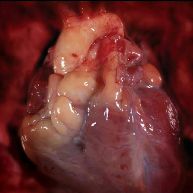
So the secret I am talking about is just a major thoracic mass of tissues that is guarded by lungs and is arranged beautifully in a cage behind our chest. Yes, it’s our heart. A HUMAN HEART.
I guess you are wondering why I called heart a secret? We all have hearts. Placed at mid-thoracic cavity tilting its apex to left side of the body, pumps blood, keep beating, and so on. Everything is known then what’s the secret is about?
Okay!!
Normally we all are aware of the vital functions and position of our heart. But do you know the different possibilities in position, function, and many more parts of the heart?
Cross-section of the human heart
OUTLOOK
The Human Heart is the first forming organ during the development process of the body. It is a muscular organ made up of the myocardium (cardiac /heart) muscle. These are involuntary, striated muscle which contributes to the main tissue of the wall of the heart.
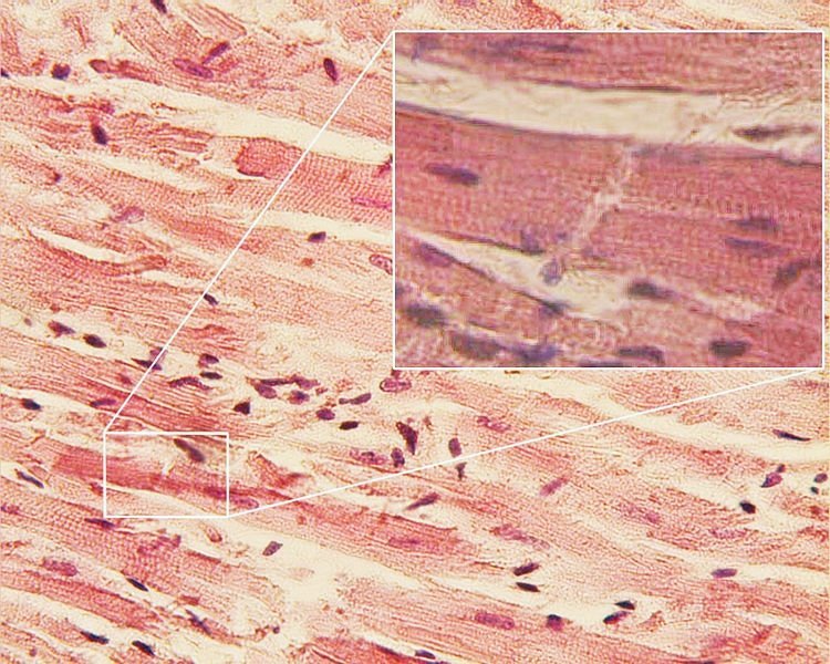
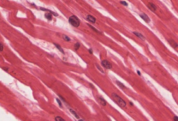
These muscles are composed of individual heart muscle cells, cardiomyocytes, joined together by intercalated discs that are encased by collagen fibers and other substances that form the extracellular matrix.
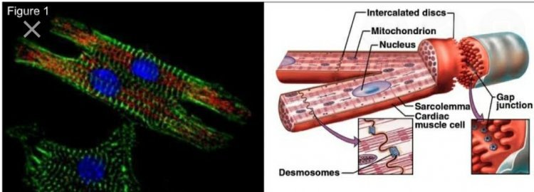
Cardiomyocytes
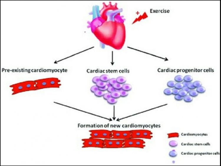
Formation of Cardiomyocytes
Location: The heart is located in the middle compartment of the chest between the lungs behind the sternum(breastbone) and above the diaphragm. The Center is located about 1.5cm to the left of the midsagittal plane. The aortic arch lies behind the heart. The esophagus and spine lie behind the heart.
Position: The heart is shaped like an upside-down pear with the upper side as a base and the lower side as the apex. The base is present in the middle of the chest and the apex is slightly tilted towards the left.
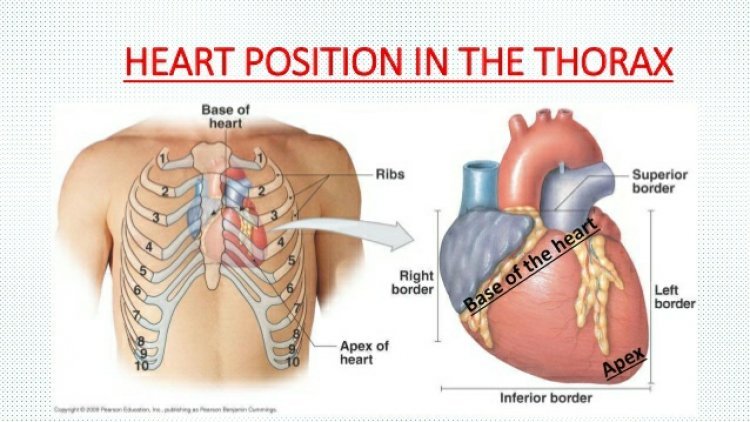
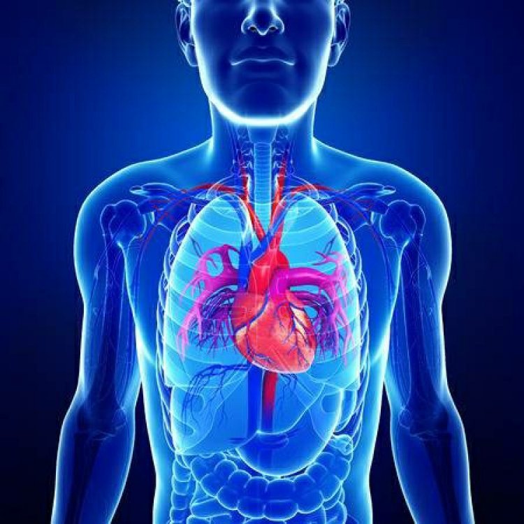
Position of heart in the human body
The size of the human heart is nearly equal to the closed fist of our hand. The average weight of an adult human heart is about 280gm-340gm. The following chart shows the weight of the heart at different ages in humans.
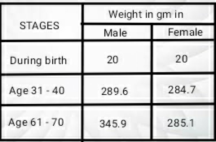
During the fetal stage weight of the heart is 20gms which occupies about 0.66 – 0.8 % of total body weight.
WHY HEART IS A VITAL ORGAN?
- To be active and functional, tissue needs a constant supply of oxygen and nutrition.
- Pumped blood carries oxygen and nutrients to the body while carrying metabolic waste(carbon dioxide) to the lungs.
- The oxygenated blood is therefore necessarily supplied to all parts of the body.
- This blood is supplied by the heart via the aorta to all parts of the body.
- Due to some dysfunctions or blockages if the heart is not able to supply blood then the tissues will collapse leading to the death of the body.
- So to keep the body alive heart is necessary. Any damage to the heart can lead to many major issues in the body.
ANATOMY
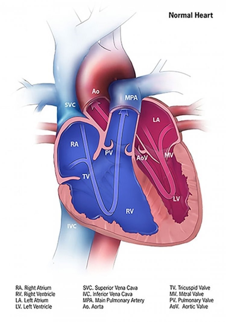
The human heart is divided into two sides: right and left side. Each side is divided into two chambers: the top chamber, and the bottom chamber. Atria are present in the base (top) and ventricles are on the apex (bottom).
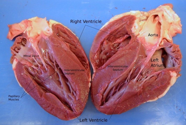
Anatomy of heart
The septum is the wall of muscles that separates the right atrium and right ventricle from the left atrium and left ventricle.
The heart is encased in a thin double-walled sac that protects and anchors the heart. This sac is called the pericardium. The outer layer of the pericardium is called as parietal pericardium layer and the serous pericardium layer is the inner one.
The pericardial fluid lubricates the heart during the contraction and movements of the lungs and diaphragm and is present in between the layers of the pericardium.
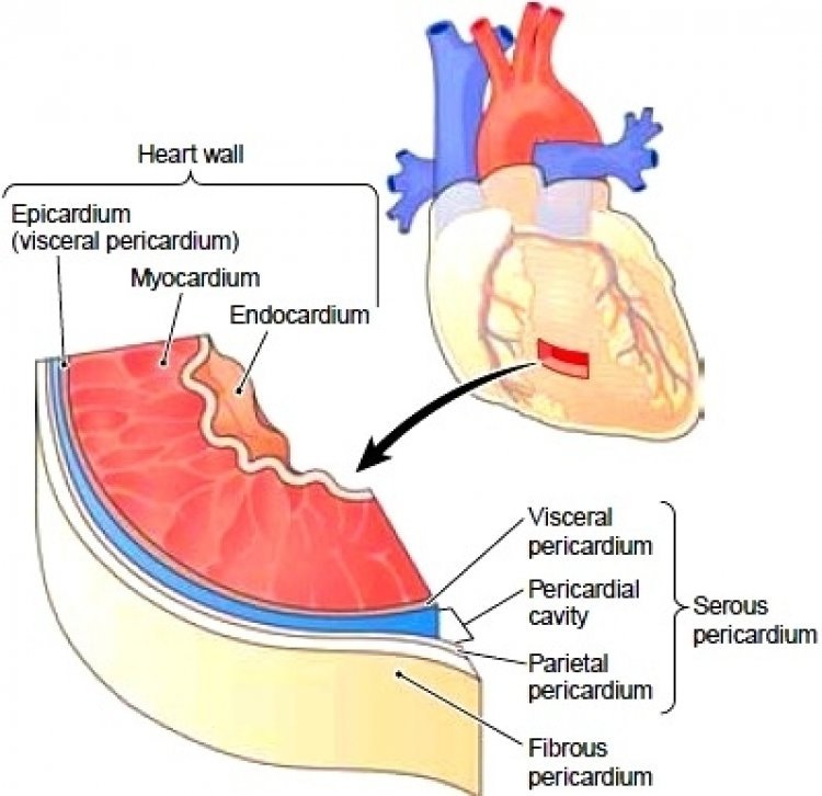
The outer wall of the heart is made up of three layers: epicardium, myocardium, and endocardium.
Epicardium is the outermost layer of the heart and at the same time, it is the last layer of the pericardium. The middle layer, the myocardium contains the muscles which contract during the cardiac cycle. The last layer is the endocardium which has a lining that has direct contact with blood.
We know that the septum separates the heart into two parts i.e. right and left. Each of them has one atrium and one ventricle. Now the atrium and ventricle are separated from each other by atrioventricular valves. The tricuspid atrioventricular valve separates the right atrium from the right ventricle and the bicuspid or mitral atrioventricular valve separates the left atrium from the left ventricle.
Valve: a structure that ensures a flow of blood in one direction only and is composed of connective tissue and endocardium.
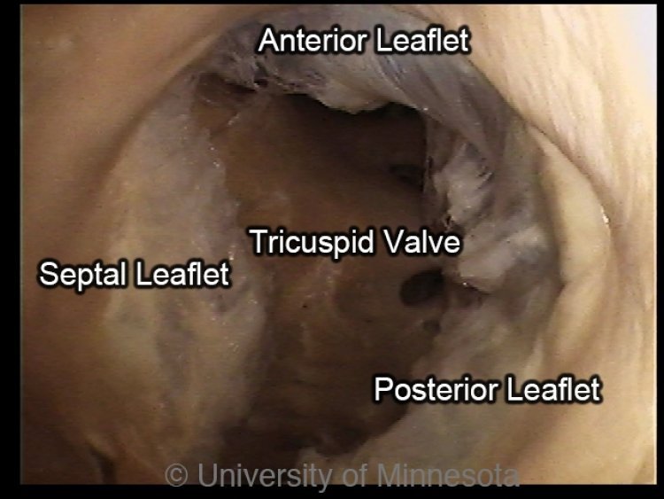
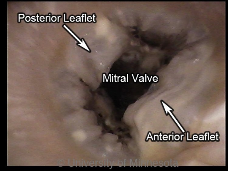
The tricuspid valve consists of three cusps: anterior, septal, and posterior.
The bicuspid valve has two cusps: anterior and posterior.
The base of each cusp is anchored to a fibrous ring that surrounds the orifice (valve which controls the blood flow).
The attachment of fibrous cords that supports mitral and tricuspid valves to the free edges of valve cups is known as chordae tendineae.
Chordae tendineae are tendons resembling fibrous cords of connective tissue which are made up of 80% of collagen and 20% of elastin fibers (approx). Chordae tendineae are also called heartstrings or tendinous chords.
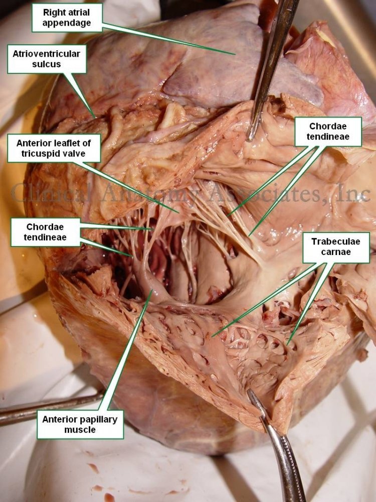
Papillary muscles are connected to atrioventricular valves via Chordae tendineae. The interior surface of the ventricles is covered with papillary muscles. These muscles contract ventricular systole to prevent the prolapse of the valve leaflets into the atria.
There are five papillary muscles in total. Three of which are present in the right ventricle and support the tricuspid valve. The remaining two are present within the left ventricle and act on the mitral valve.
Semilunar valves are located in between the ventricles and outflow vessels. In between the right ventricle and the pulmonary trunk pulmonary valve is located. This valve consists of three cusps: left, right, and anterior.
The aortic valve is located in between the left ventricle and ascending aorta. This aortic valve consists of three cusps: right, left, and posterior.
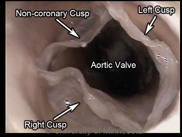
Pulmonary and aortic valves have a similar structure. The sides of each valve leaflet are attached to the walls of the outflow vessel, which is slightly dilated to form a sinus. The free superior edge of each leaflet is thickened (lunule) and is widest in the midline (nodule).
Sinoatrial (SA) Node: It is a specialized myocardial structure located on the atrial wall at the junction of the superior caval vein and right atrium. SA produces an electrical pulse that derives heart contractions.
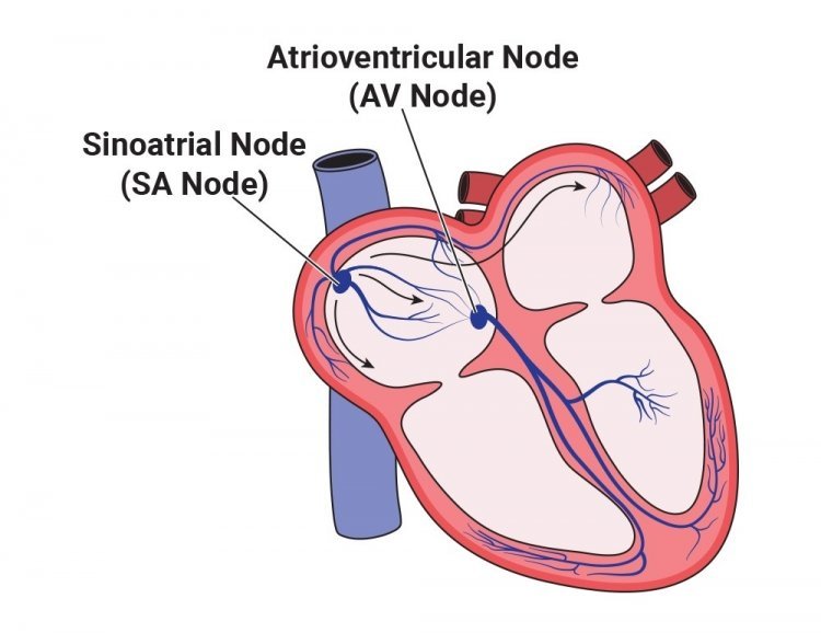
Atrioventricular (AV) Node: It is the electric relay station between the upper and lower chamber of the heart. Located in the Koch triangle near the coronary sinus on the interatrial septum. AV node controls heart rate and is the major element in the cardiac conduction system.
✧・✧・✧・✧


